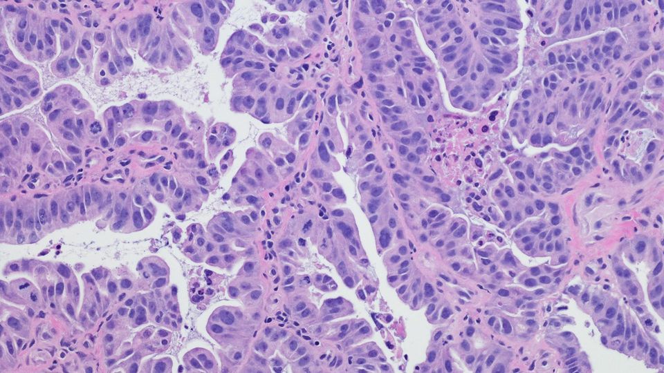
Considering the spatial distribution of immune cells is crucial when determining the prognosis and overall survival outcomes of cancer patients. Nevertheless, despite the valuable transcriptomic information provided by probe-based spatial assays, detecting immune cell infiltration can pose challenges due to their low mRNA content.
This poster highlights assay solutions that enable the spatial characterization of immune cell populations within multiple tissue types, including immune-rich human tonsil, lymph node and breast cancer.
Download this poster to learn more about:
• Conducting whole-transcriptome analyses with human FFPE tissues
• Achieving high-quality transcriptomic and proteomic libraries with a spatial biology context
• Highly specific and sensitive immune cell epitope detection
Proteogenomic characterization of individual cores in the breast cancer TMA with Visium CytAssist
We characterized the performance of the Visium CytAssist Gene and Protein
Expression assay on immune-rich human tonsil, lymph node, and breast cancer
tissue. FFPE tonsil and lymph node sections were H&E stained and FFPE
breast cancer sections were IF stained. After staining, these samples went
through the workflow described in Figure 1 using the 6.5 mm capture area slide.
The breast cancer tissue microarray (TMA) (NBP2-42060, Novus Biologicals)
was constructed using samples from 64 breast cancer patients. Prior to the
main workflow, the TMA slide was H&E stained and relevant morphological
features identified. Bottom-right 24 cores were selected for further
transcriptomic and proteomic analyses (dotted square, Fig. 3A). Gene and
protein libraries generated were sequenced on Illumina NovaSeq sequencer as
per 10x Genomics’ recommendations: at least 25k reads per spot (rps) for gene
libraries, and 5k rps for protein libraries.
Spatially resolved whole-transcriptome analysis with multiplexed protein
detection in human FFPE tissues
Anushka Gupta, Nancy Conejo, Rena Chan, Ace Santiago, Anuj Patel, Hardeep Singh, Stephen Williams, Govinda
Kamath, David Sukovich, Augusto Tentori
10x Genomics, Pleasanton, CA, USA, 94588
The tumor microenvironment is composed of highly heterogeneous niches, often with
varying degrees of immune infiltration. The spatial distribution of immune cells with
respect to malignant cells can directly impact patient prognosis and overall survival
outcomes. The Visium CytAssist Spatial Gene Expression assay uses a whole
transcriptome, probe-based approach to detect and quantify mRNA expression with
spatial context. Although examination of the tumor microenvironment with a
probe-based spatial assay can provide significant transcriptomic information
concerning regions of interest, immune cells frequently have low mRNA content (~25x
lower than a typical mammalian cell1
) and can be difficult to detect. The use of
antibody-conjugated probes specific to immune cell epitopes, which have higher
expression counts than their transcripts2
, can enhance data recovered from these
tumor samples, enabling spatially accurate detection of immune cells. The Visium
CytAssist Gene and Protein Expression assay enables identification of immune-specific
epitopes via a panel of 31 antibodies, each of which is conjugated to a DNA probe.
Protein-level measurements are derived from the same FFPE tissue section used for
whole-transcriptome analysis. Using the CytAssist workflow, we showcase the ability to
identify immune cell localization within multiple immune and tumor tissues, including an
array of human breast cancer punches. Spatial expression patterns of immune markers
map back to distinct morphological features within the samples. Overall, these data
highlight the value of Visium CytAssist Spatial Gene and Protein Expression assay in
immuno-oncology studies, through the integration of spatially resolved transcriptomic
and immune cell protein marker data for FFPE tissue sections.
Abstract
Visium CytAssist Gene and Protein Expression
Workflow
Visium CytAssist Gene and Protein Expression
Capabilities
Experimental Design
11mm Slide
Figure 1. An overview of the Visium CytAssist Gene and Protein Expression workflow
● Simultaneous transcriptome and immune cell epitope detection from human FFPE
tissue sections mounted on standard glass slides
● Compatible with Hematoxylin & Eosin (H&E) and immunofluorescence (IF) staining
● Flexibility to select target regions for analysis based on morphological or
protein-level landmarks of interest
● Probe-based chemistry for transcriptomic analysis and antibody-conjugated probes
for proteomic analysis
● Two Capture Areas per slide with spatially barcoded spots
● Two slide formats: 6.5 x 6.5 mm or 11 x 11 mm Capture Areas with either ~5,000
(6.5 mm) or ~14,000 (11 mm) uniquely barcoded spots
● Each barcoded spot has capture sequences for RTL probes (poly-dT) and
antibody-conjugated probes
● 31 antibodies in the panel focused on various immune markers, including both cell
surface and intracellular proteins
● 4 isotype controls for assessing non-specific antibody binding, and for normalization
of proteomic UMI counts
Spatial Gene and Protein Expression Profiling of Immune and Cancer Tissues with Visium CytAssist
Figure 2. Gene and Protein Expression slide with spatially-barcoded capture areas
Figure 3. H&E-stained human breast cancer TMA on standard slide. (A) TMA is constructed
using samples from breast cancer patients, 1 placenta control (A1), and 1 negative paraffin
control (A2, dotted circle) (B) Patient information from the region of analyte transfer depicted as
grid position, tissue type, patient age, TMN staging, and cancer staging
5 4 3 2 1
A
B
C
D
E
(A) F
(B)
Conclusions and Acknowledgements
Figure 4. (A) H&E-stained human tonsil and lymph node sections (left). Isotype-normalized,
log-transformed counts for Cd3e (T-cell marker) and Pax5 (B-cell marker) markers at the
protein- and gene-level (B) Distribution of total UMI counts per spot for the
antibody-conjugated probe panel, and distribution of total genes detected per spot, in tonsil
and lymph node. Note that Visium CytAssist Gene and Protein Expression assay
detects ~1M Antibody-UMIs, while also detecting ~6000 genes for every 55um-sized
spot (C) DAPI-stained human breast cancer section (left). Expression pattern for Pcna and
Vim protein as visualised using IF staining, or using the Visium CytAssist workflow, r
represents the correlation between IF and Cytassist protein expression profiles.
Figure 5. (A) H&E-stained human breast cancer tissue microarray on standard slide (left).
Expression of ubiquitously expressed immune marker protein Cd4 in the TMA (center). Variable
expression of protein PanCK across different cores in the TMA (right) (B) Pathologist annotated
H&E staining of core D5 (left, dotted square in A). Expression of proteins Bcl5 (associated with
plasma B cells), and Pcna (associated with cellular proliferation) in core D5 (C) H&E staining of
core E2 (left, dotted square in A). Expression of proteins Bcl5, and Pcna in core E2. Expression for
each protein is plotted as isotype-normalized, log-transformed counts. Tonsil
H&E Cd3e (Protein) CD3E (Gene) Pax5 (Protein) PAX5 (Gene)
Lymph Node
Breast Cancer
IF Stain
Cytassist (Protein)
Pcna Vim
H&E Cd4 (Protein) PanCK (Protein)
Necrosis
Invasive Carcinoma
H&E D5 H&E E2
Bcl2 (Protein) Pcna (Protein)
2.0 mm
0.5 mm
0.5 mm
The Visium CytAssist Gene and Protein Expression assay from 10x Genomics enables whole-transcriptome analysis with multiplexed immune cell epitope detection in
human FFPE tissues. Spatial expression profile for proteins derived using the Visium CytAssist agrees well with IF staining. Immune cell epitope detection using
antibody-conjugated probes does not interfere with transcript capture, and the overall workflow yields high-quality transcriptomic and proteomic libraries in a spatial
context. Immune cell epitope detection is highly specific and sensitive, as demonstrated by mapping of immune markers to distinct morphological features within the
samples. Here we focus our analyses on human tonsil, lymph node, and breast cancer, although, multiple other human tissue types are compatible with the Visium
CytAssist Gene and Protein Expression workflow by following 10x Genomics recommended sample preparation and staining protocols.
Thanks to the Visium R&D, TechComm, and Legal Team at 10x Genomics Headquarters, Pleasanton, CA. Special thanks to Areeb Mallick and Syrus Mohabbat for
assistance with sample preparation and tissue sectioning. For more information, please contact: info@10xgenomics.com
1. J Racle et al. (2017), Simultaneous enumeration of cancer and immune cell types from bulk tumor gene expression data, eLife 6:e26476.
2. J Li et al. (2020), Discrepant mRNA and Protein Expression in Immune Cells., Curr Genomics. 21(8):560-563.
3.0 mm
1.5 mm
Sensitivity Metric: Tonsil Sensitivity Metric: Lymph Node
(A)
(B)
(C)
2.4 mm
2 mm
Pax5: B-cell Marker
Cd3e: T-cell Marker
(A) (B)
(C)
IF or H&E Stain Probe Release
r=0.8
r=0.8
Iso-norm Log-norm Iso-norm Log-norm
Iso-norm Log-norm Iso-norm Log-norm
Iso-norm
Iso-norm
Iso-norm Iso-norm
Iso-norm Iso-norm
Iso-norm Iso-norm


