GC-MS Principle, Instrument and Analyses and GC-MS/MS
Complete the form below to unlock access to ALL audio articles.
What is gas chromatography mass spectrometry?
Gas chromatography mass spectrometry (GC-MS) consists of two very different analytical techniques: gas chromatography (GC) which is hyphenated (hence uses a hyphen not a forward slash) to mass spectrometry (MS). Usually, the analytical instrument consists of a gas chromatograph that is hyphenated via a heated transfer line to the mass spectrometer, and the two techniques take place in series. However, some specialist and usually miniature or portable instruments contain the whole GC-MS within a single box.
How does a GC-MS instrument work?
GC-MS analysis and what the retention time tells you
GC is a separation science technique that is used to separate the chemical components of a sample mixture and then detect them to determine their presence or absence and/or how much is present. GC detectors are limited in the information that they give; this is usually two-dimensional giving the retention time on the analytical column and the detector response. Identification is based on comparison of the retention time of the peaks in a sample to those from standards of known compounds, analyzed using the same method. However, GC alone cannot be used for the identification of unknowns, which is where hyphenation to an MS works very well. MS can be used as a sole detector, or the column effluent can be split between the MS and GC detector(s).
MS is an analytical technique that measures the mass-to-charge ratio (m/z) of charged particles and therefore can be used to determine the molecular weight and elemental composition, as well as elucidating the chemical structures of molecules. Data from a GC-MS is three-dimensional, providing mass spectra that can be used for identity confirmation or to identify unknown compounds plus the chromatogram that can be used for qualitative and quantitative analysis.
How does a GC-MS instrument work?
The sample mixture is first separated by the GC before the analyte molecules are eluted into the MS for detection.1 They are transported by the carrier gas (Figure 1 (1)), which continuously flows through the GC and into the MS, where it is evacuated by the vacuum system (6).
1. The sample is first introduced into the GC manually or by an autosampler (Figure 1 (2)) and enters the carrier gas via the GC inlet (Figure 1 (3)). If the sample is in the liquid form, it is vaporized in the heated GC inlet and the sample vapor is transferred to the analytical column (Figure 1 (4)).
2. The sample components, the “analytes”, are separated by their differences in partitioning between the mobile phase (carrier gas) and the liquid stationary phase (held within the column), or for more volatile gases their adsorption by a solid stationary phase. In GC-MS analyses, a liquid stationary phase held within a narrow (0.1-0.25 mm internal diameter) and short (10-30 m length) column is most common.
3. After separation, which for GC-MS analyses doesn’t require total baseline resolution unless the analytes are isomers, the neutral molecules elute through a heated transfer line (Figure 1 (5)) into the mass spectrometer.
4. Within the mass spectrometer, the neutral molecules are first ionized, most commonly by electron ionization (EI). In EI, an electron, produced by a filament, is accelerated with 70 electron volts (eV) and knocks an electron out of the molecule to produce a molecular ion that is a radical cation. This high energy ionization can result in an unstable molecular ion and excess energy can be lost through fragmentation. Bond breakage(s) can lead to the loss of a radical or neutral molecule and molecular rearrangements can also occur. This all results in a, sometimes very large, number of ions of different masses, the heaviest being the molecular ion with fragment ions of various lower masses, depending on:
- the molecular formula
- the molecular structure of the analyte
- where bond breakage has occurred
- which part has retained the charge
5. The next step is to separate the ions of different masses, which is achieved based on their m/z by the mass analyzer (Figure 1 (8)).
There are numerous different mass analyzer types, and this is where the vast differences in mass resolution (and hence instrument price) is seen. Mass resolution is the ability of the mass analyzer to separate ions with very small differences in m/z. Unit mass resolution instruments can only separate nominal masses or those down to a single decimal place, whereas high mass resolution (HRMS) instruments can separate them to four or five decimal places.
The most common type of unit mass instrument is the quadrupole, which is a scanning instrument and varies the voltage to allow only ions of a certain m/z to have a stable trajectory through the four poles to reach the ion detector. Quadrupole instruments are used in two different modes of operation:
- Full scan mode, where all ions are acquired across a mass range, useful for identification of unknowns, method development and qualitative and quantitative analysis for higher concentration analytes.
- Selected ion monitoring (SIM) mode, where only selected ions that represent the target compound are acquired, useful for trace analysis, as higher sensitivity is obtained, but only of target analytes.
An ion trap is also a scanning instrument but is three-dimensional, trapping the ions in mass-dependent orbits before ejecting them sequentially to reach the ion detector.
Time-of-flight (ToF) mass analyzers separate the ions based on the time they take to travel down the flight tube to reach the ion detector. With the same kinetic energy, those with lower masses have a higher velocity and therefore arrive first, whereas those with higher masses have a lower velocity and arrive later. ToFMS instruments can range in mass resolution and acquisition rate: very fast ToFs, with acquisition rates of up to 1000 spectra/second are unit mass resolution, whereas HRMS ToFs have a lower acquisition rate. High acquisition rates are good for two-dimensional GC (GC x GC) applications with peak widths down to 30 ms, however HRMS is very useful to determine the molecular formula. Therefore, there are ToFs on the market that range in speed and mass resolution, the choice of which is dependent on the application, but the GC peak width must match the acquisition rate capabilities of the MS.
Other HRMS instruments that are hyphenated to GC include the magnetic sector mass analyzer, which bends the trajectories of the ions to separate them using electric and magnetic fields. Magnetic sector GC-MS instruments are more commonly found in isotope ratio analyses.
In the HRMS orbitrap, the ions orbit around a central spindle and the frequency that they move up and down the central spindle is m/z-dependent.
6. After the ions have been separated by the mass analyzer based on their m/z, they reach the ion detector (Figure 1 (9)) where the signal is amplified by an electron multiplier (for most low resolution MS) or a multi-channel plate (for most HRMS instruments). The signal is recorded by the acquisition software on a computer (Figure 1 (10)) to produce a chromatogram and a mass spectrum for each data point.
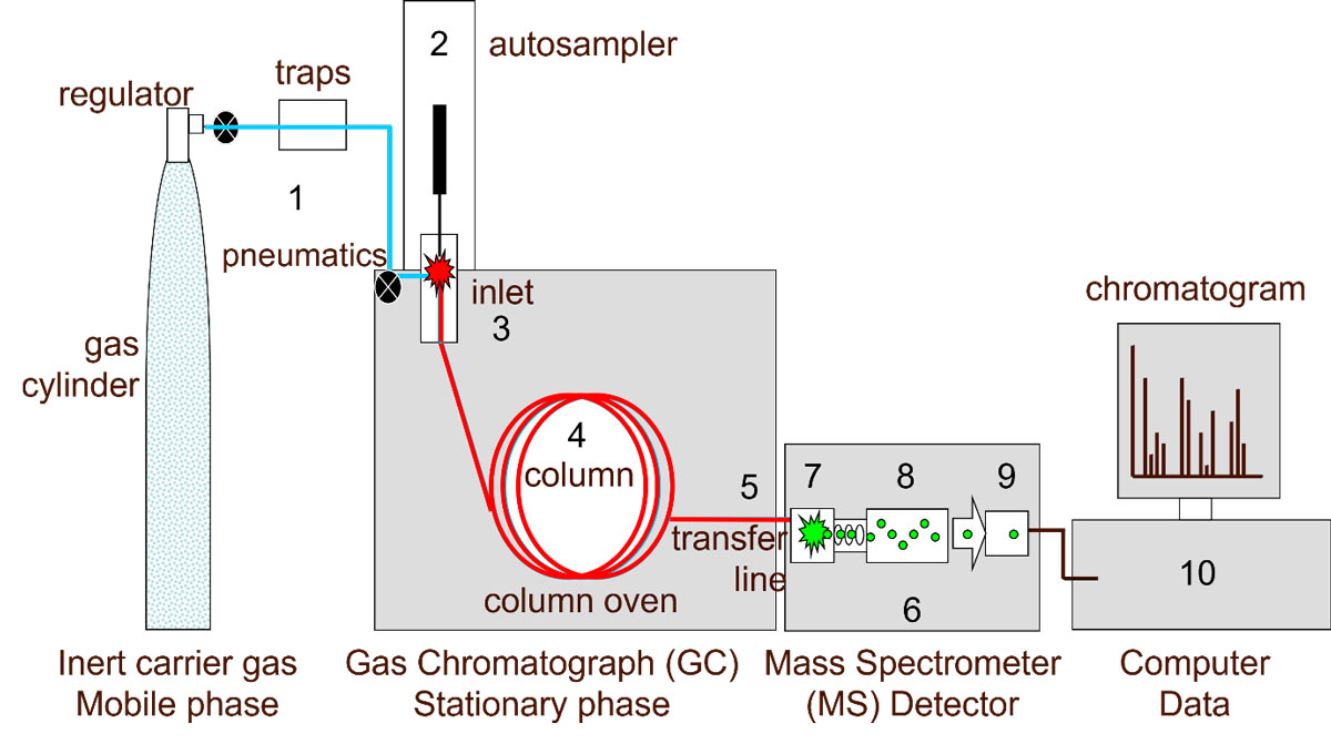
Figure 1: A simplified diagram of a gas chromatograph–mass spectrometer showing (1) carrier gas, (2) autosampler, (3) inlet, (4) analytical column, (5) interface, (6) vacuum, (7) ion source, (8) mass analyzer, (9) ion detector and (10) PC. Credit: Anthias Consulting.
GC-MS analysis and what the retention time tells you
GC-MS data is three-dimensional, as shown in Figure 2. The x-axis shows the retention time; the time from sample injection to the end of the GC run. This can also be viewed as the scan number, which is the number of data points that have been acquired by the MS across the run. The y-axis is the response or intensity measured by the ion detector (Figure 1 (9)). The z-axis is the m/z of the ions across the mass range acquired.
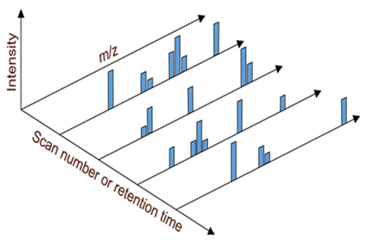
Figure 2: GC-MS data is three-dimensional, giving scan number/retention time, response/intensity and m/z. Credit: Anthias Consulting.
The two-dimensional chromatogram, as shown in Figure 3, is produced by summing the abundances of all the ions at a single data point and plotting it against the retention time (RT)/scan number to produce a total ion chromatogram (TIC), which is more comparable to a chromatogram produced by a GC detector. However, each data point in the total ion chromatogram is a separate mass spectrum and can usually be opened in a separate window in the software. In the example shown in Figure 3, the apex data point of peak 3 has been opened.
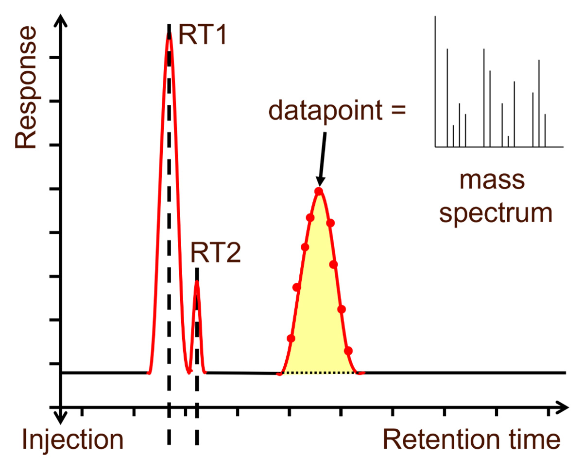
Figure 3: Total ion chromatogram (TIC) output from a GC-MS. Credit: Anthias Consulting.
An example mass spectrum of the straight chain hydrocarbon n-decane can be seen in Figure 4. The molecular ion, 142 m/z can be seen on the far right. As decane is a saturated hydrocarbon, the excess energy from ionization cannot be delocalized internally and therefore most molecular ions fragment, resulting in many fragment ions and a low abundance of the molecular ion. The longer the chain length of a saturated hydrocarbon, the lower the abundance of the molecular ion until no molecular ion is observed in the mass spectrum. However, unsaturated molecular ions and especially those with conjugated double bonds, like aromatic compounds, have less fragmentation as the excess energy can be internalized more easily. Another observation from the mass spectrum of decane is the series of fragment ions at m/z 43, 57, 71, 85, 99 and 113 that differ by an m/z of 14. These are formed by the overlapping cleavage of bonds at successive -C2H4- units, if the charge is +1 this equates to a mass of 28 unified atomic mass units (u) and is a key feature of mass spectra from hydrocarbons. The mass spectrum is a fingerprint of the molecule and, if obtained using the same ionization technique and voltage, can be compared to libraries of spectra obtained using the same technique at the same voltage. The most common commercial libraries are EI spectra produced at 70 eV. The mass spectrum can also be interpreted to determine the molecular formula and structure of the molecule using the masses of the ions, the presence of isotopes and the losses between the fragment ions.
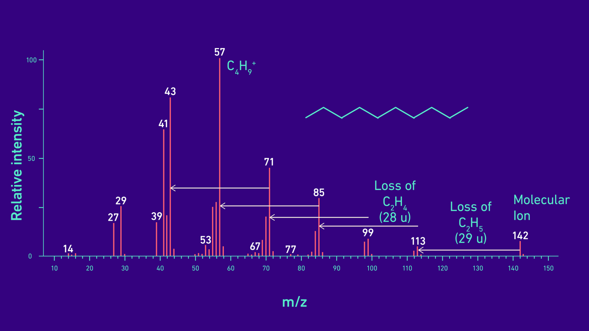
Figure 4: Example mass spectrum of decane (C10H22), a straight chain hydrocarbon.
In GC, the retention time is used for the identification of target analytes and usually the area is used for quantitation. For accurate quantitation, the peaks need to have good chromatographic separation with baseline resolution, as shown in Figure 3 peaks at RT1 and RT2. With GC-MS the mass spectrum provides an additional method for confirmation of the target analyte using the full mass spectrum or using the presence and relative ratios of a few of the ions. Quantitation using GC-MS data is usually from the area of a single, unique ion as it is less likely to have interferences from co-eluting peaks than by using the area under the TIC peak. As such, chromatographic baseline resolution isn’t needed for accurate quantitation, as long as a unique ion can be selected that isn’t present in the coeluting peaks and therefore the peaks are spectrally resolved, and baseline-to-baseline integration can be achieved.
GC-MS vs GC-MS/MS
The use of chromatographic and spectral resolution is very powerful for the separation and identification of target analytes. However, in the analysis of trace analytes down to femtogram (fg) levels in very complex samples, for example environmental, food or biological samples, the matrix can be overwhelming.2, 3 Sample preparation can be used to remove the majority of the matrix; however, analyte molecules are also frequently lost. Chromatographically, the matrix peaks can be separated from analyte peaks using GC x GC, where two columns with different stationary phases are used. Within the mass spectrometer, spectral resolution can be used, where unique ions for the target analyte are chosen that aren’t present in the coeluting matrix peaks. However, this frequently fails when analyzing these very complex samples, as fragment ions from the various coeluting matrix peaks have the same m/z as many ions from the target peak, skewing the ion ratios and resulting in false negatives or inaccurate quantitation.
Tandem mass spectrometry (MS/MS) uses multiple stages (mass analyzers) within the mass spectrometer to increase the sensitivity of the analyte by reducing the background from the coeluting matrix peaks. Different, chromatographically coeluting compounds may create molecular/fragment ions that have the same m/z; however, these ions will have a different structure. After ionization, the first mass analyzer selects the ion, called the precursor ion. The next stage is to fragment this further, which is usually achieved with an inert gas (e.g., argon) through a process called collision induced dissociation (CID). The ions produced, called product ions, will be dependent on the structure of the precursor ion, therefore the product ion mass spectrum from the interferent(s) will be different from that of the target analyte. The next stage of MS/MS is then to separate the product ions using another mass analyzer before the ion detector. There are several different configurations of MS/MS instruments including a triple quadrupole (QqQ) where Q1 is used for precursor selection, q2 is used as the collision cell and Q3 is used as the product ion mass analyzer. Triple quadrupoles are the most common as they are unit mass instruments and therefore more affordable. An example of a triple quadrupole is shown in Figure 5 in multiple reaction monitoring (MRM) mode. Q-ToFs use a quadrupole for precursor ion selection and an HRMS ToF as the product ion mass analyzer. Ion traps are capable of performing MSn, with all stages occurring within the single trap. All ions are ejected except for the precursor ions and a voltage is applied that resonates with the energy to destabilize and fragment the precursor ion of that particular m/z. This process can then be repeated to fragment further, as many times as are needed.
Generally, MS/MS instruments are used for target analysis only and not for the routine analysis of unknowns. There are several modes of operation including MRM, single reaction monitoring (SRM) which is similar but more sensitive, product ion scans where Q3 is operated in scan mode to acquire the full product ion mass spectrum and neutral loss scans. MS/MS instruments can also be used as a standard MS—this is required during method development of MS/MS techniques—however, caution should be applied as the mass spectrum produced may be of a lower quality and affect the identification, particularly for scanning instruments. Some manufacturers also enable dual acquisition mode, where the acquisition alternates between e.g., MRM mode for target analysis and scan mode for the identification of unknowns.

Figure 5: An example of a triple quadrupole MS/MS instrument in multiple reaction monitoring (MRM) mode. Credit: Anthias Consulting.
Strengths and limitations of GC-MS
GC alone is limited in that it isn’t possible to identify unknown compounds using standard GC detectors, but this is possible when paired with MS. Conversely, direct analysis of samples using MS produces mixed mass spectra that can be difficult to deconvolute and interpret, especially when there are more than a few compounds in the sample. But pairing with GC gives the ability to separate the mixture.
Not all compounds can be separated on a standard GC column, therefore the ability to use spectral resolution to remove the interferences leads to more accurate quantitation. The deconvolution of mass spectra enables the detection of small peaks under baselines and matrix peaks, and selective MS/MS enables the identification of trace analytes in very complex samples4 with little interference.
Isomers of an analyte usually have the same or very similar mass spectra and therefore it is very difficult to distinguish between them using just the mass spectrum. Therefore, chromatographic resolution is required to separate them. The individual isomers are identified by their retention times and good chromatographic resolution is required for accurate integration and quantitation. This is the same for homologous series, such as saturated hydrocarbons, where beyond a certain chain length, no molecular ion can be seen and the mass spectra all look the same. Again, the retention time must be used to determine the identity of the saturated hydrocarbon.
In terms of the lack of a molecular ion used in the identification of a compound, alternative softer ionization methods can be used. For example, EI at a lower eV or chemical ionization in positive or negative modes can result in fewer fragment ions and prevent the loss of the molecular ion or make its peak stronger. However, the fragmentation pattern is very much used for identification of the compound’s structure, and target libraries usually need to be created as few exist for soft ionization techniques, but this is improving as soft ionization techniques become more extensively used.
The hyphenation of GC and MS brings together two powerful techniques that are complementary to each other and that can be used for the separation and identification of compounds in a sample. When one method fails to provide answers, the other method can then be relied upon.
Common problems with GC-MS
The most common problems in GC-MS instruments are leaks into the system and system contamination.
The MS usually has a vacuum to reduce background interferences and ion-molecular reactions, to increase the lifetime of components, reduce maintenance and to avoid any electrical discharges, as high voltages are used within the MS. A very good vacuum is required to obtain good sensitivity and small leaks can increase the background whereas large leaks impede the vacuum system. High column flows increase the number of gas molecules entering the MS and the vacuum system cannot evacuate them fast enough to establish a good vacuum, so sensitivity reduces. Hence, when moving a method from GC to GC-MS, smaller internal diameter columns should be used with lower column flows. The maximum flow rate for high sensitivity analyses is dependent on the vacuum system of the mass spectrometer. This should be known and taken into consideration, along with the optimal flow rate of the carrier gas, for the best separation efficiency.
Mass spectrometers are sensitive instruments and regular maintenance is required to clean the ion source and replace the oil in the vacuum pump to retain good sensitivity. High molecular weight matrix from the sample should be retained in the GC inlet liner, by optimizing the inlet temperature, which is easily replaced rather than transferring into the column where it can damage the stationary phase and dirty the MS ion source. Another source of ion source contamination is column bleed. More stable “-MS” columns should be installed in a mass spectrometer and column conditioning should not be carried out when connected to the MS.
GC-related issues still occur, for example activity and sample degradation, especially where the sample has been through less sample preparation, for example if GC-MS/MS is used and the matrix can’t be “seen”.
Applications of GC-MS and GC-MS drug testing
GC-MS and GC-MS/MS are used in many industries for routine analysis looking for volatile contaminants with a molecular weight of usually less than 700 amu, for example in the food,2 environmental,5 forensics,6 anti-doping7 and consumer products8 industries. GC-MS is also used in research to identify unknown volatile compounds, including in food and flavors, space, petrochemical, chemical, agriculture, tobacco, pharmaceutical, healthcare, energy, mining, environmental and forensics to name but a few.
Drug testing, as an example, can be carried out for multiple reasons: pathology, healthcare and anti-doping of both humans and animals.9 Biological fluids like urine and blood samples are complex with large amounts of matrix and usually the drugs are at very low concentration. Therefore, their detection requires a hyphenated technique like GC-MS to start to separate the target or compounds of interest away from the matrix peaks. They can then be selectively identified using MS/MS and more frequently MS/MS with an HRMS and sometimes additional GC x GC separation.
References
1. Turner DC, Schäfer M, Lancaster S, Janmohamed I, Gachanja A, Creasey J. Gas Chromatography–Mass Spectrometry. Royal Society of Chemistry; 2020. ISBN-10:1782629289
2. Hernández F, Cervera MI, Portolés T, Beltrán J, Pitarch E. The role of GC-MS/MS with triple quadrupole in pesticide residue analysis in food and the environment. Anal Methods. 2013;5(21):5875-5894. doi:10.1039/C3AY41104D
3. Fialkov AB, Steiner U, Lehotay SJ, Amirav A. Sensitivity and noise in GC–MS: Achieving low limits of detection for difficult analytes. Int. J. Mass Spectrom. 2007;260(1):31-48. doi:10.1016/j.ijms.2006.07.002
4. Bünning TH, Strehse JS, Hollmann AC, Bötticher T, Maser E. A toolbox for the determination of nitroaromatic explosives in marine water, sediment, and biota samples on femtogram levels by GC-MS/MS. Toxics. 2021;9(3):60. doi:10.3390/toxics9030060
5. Ferrer I, Thurman EM. Advanced Techniques in Gas Chromatography-Mass Spectrometry (GC-MS-MS and GC-TOF-MS) for Environmental Chemistry: Volume 61. Elsevier; 2013;2-502 ISBN:9780444626240
6. Pasternak Z, Avissar YY, Ehila F, Grafit A. Automatic detection and classification of ignitable liquids from GC–MS data of casework samples in forensic fire-debris analysis. Forensic Chem. 2022;29:100419. doi:10.1016/j.forc.2022.100419
7. Narciso J, Luz S, Bettencourt da Silva R. Assessment of the quality of doping substances identification in urine by GC/MS/MS. Anal Chem. 2019;91(10):6638-6644. doi:10.1021/acs.analchem.9b00560
8. Karlberg AT, Albadr MH, Nilsson U. Tracing colophonium in consumer products. Contact Derm. 2021;85(6):671-678. doi:10.1111/cod.13944
9. Clarke J, Rockwood AL, Kushnir MM. Mass spectrometry. In: Rifai N, Horvath AR, Wittwer CT, Hoofnagle A ed. Principles and Applications of Clinical Mass Spectrometry: Small Molecules, Peptides, and Pathogens. 1st ed.; Elsevier; 2018:33-65. ISBN:9780128160633
About the author:
Diane is the senior consultant and director of Anthias Consulting Limited. She has developed methods, given support and high-quality training for companies in most industries around the world for more than 20 years. A Warwick University Graduate, Diane completed her Masters in analytical chemistry and started her career in environmental chemistry, later gaining significant experience as an Applications Chemist. Diane's area of research through her PhD studies at The Open University was disease diagnosis. Diane is President-Elect of the Royal Society of Chemistry Analytical Division and Chair of the Analytical Chemistry Trust Fund. Diane is a visiting academic and consultant at The Open University.


